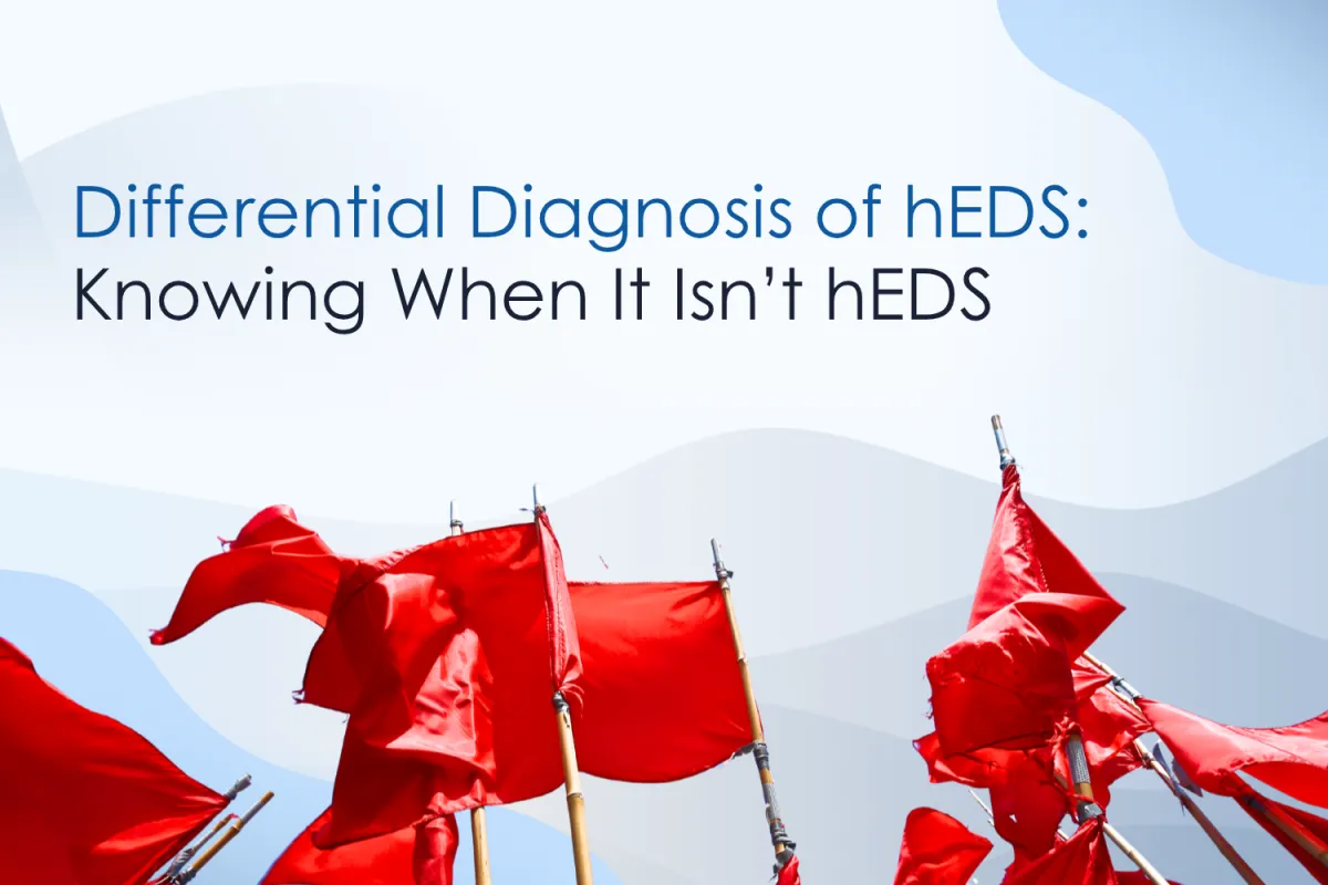
Differential Diagnosis of Hypermobile Ehlers-Danlos Syndrome: Knowing When It Isn't hEDS
Disclaimer: This resource is designed for education and general guidance only. Medical professionals are responsible for applying their own clinical judgment when using this tool. It is not a replacement for personalised medical advice. If you are seeking care, please consult a qualified medical professional for advice tailored to your situation.
Hypermobility presentations sit at the intersection of common and rare. For most clinicians working in this space, Hypermobile Ehlers-Danlos Syndrome (hEDS) and Hypermobility Spectrum Disorders (HSD) account for the majority of patients seen in practice (Castori et al., 2017; Hakim & Grahame, 2003). This familiarity is clinically valuable, but it can also create blind spots. When hypermobility becomes the default explanatory framework for every presentation, alternative diagnoses may be missed, delayed, or inappropriately dismissed.
This article is written for clinicians who encounter and recognise hypermobility but are at times concerned that they may be missing something else. The focus is not on re-teaching diagnostic criteria, but on strengthening differential diagnosis, recognising red flags, and knowing when a hypermobility presentation should prompt further medical or genetic assessment.
It also introduces a practical, red flag screening tool (FOCUSSED) that can be incorporated into routine assessment. A free printable version that's created for everyday use in clinics is available for clinicians who join the Hypermobility Project newsletter.
hEDS as a Diagnosis of Inclusion and Exclusion
HEDS remains a clinical diagnosis. By definition, the diagnosis is made after exclusion of other heritable disorders of connective tissue, particularly those associated with vascular, ocular, or organ fragility (Malfait et al., 2017). This exclusionary step is often a barrier overlooked in day-to-day practice.
In reality, many clinicians rely on pattern recognition - and understandably so. A hypermobile patient with chronic pain, fatigue, dysautonomia, and soft tissue injury history looks like hEDS because most of the time it is. The challenge is that several rare but high-risk conditions can initially present with overlapping features (Loeys & Dietz, 2024), making them difficult to distinguish in early stages or brief consultations.
Recent data from the HEDGE (Hypermobile Ehlers-Danlos Genetic Evaluation) study supports the value of systematic screening. Preliminary findings presented in November 2024 found that 7 in 1000 participants with clinically diagnosed hEDS had an identifiable alternative genetic connective tissue disorder (The Ehlers-Danlos Society, 2024). Though rare, these potentially life-threatening diagnoses can be missed without systematic red flag screening in routine clinical practice.
The consequences of missing these diagnoses extend beyond diagnostic delay. Unidentified vascular pathology can lead to arterial dissection, organ rupture, or sudden cardiac events - complications that may be preventable with appropriate monitoring and management. Early recognition allows for risk stratification, cardiovascular surveillance, activity modification, and surgical planning when indicated, fundamentally changing patient outcomes and family screening pathways.
Why "FOCUSSED" Red Flag Screening Matters
Red flag screening is not about casting a wide net or generating unnecessary anxiety. It is about systematic identification of features that fall outside the expected hEDS phenotype and that carry clinical implications.
The FOCUSSED red flag screening framework was developed and is soon to be published by rheumatologist Dr Alan Hakim in the forthcoming 9th edition of the textbook Rheumatology, a widely regarded cornerstone reference in the field (Hakim, 2026).
Dr Hakim, Chief Medical Officer at the Ehlers-Danlos Society, has been very generous in sharing his work with us and has collaborated with Sharon Hennessey to provide the downloadable resource available when signing up for the Hypermobility Project newsletter.

Sharon Hennessey & Dr. Alan Hakim.
This evidence-informed framework supports clinicians to screen efficiently and consistently during routine assessment, without turning every hypermobility consultation into a complex genetics workup. The tool is brief, practical, and designed to integrate seamlessly into existing assessment workflows. Please see the image for the key to the acronym.

The FOCUSSED acronym used to identify red flags in those with suspected heritable connective tissue disorders.
The FOCUSSED acronym prompts consideration of red flags across eight key domains:
Family history
Organs and internal structures
Cardiovascular features
Unexplained sudden death
Skeletal features
Skin findings
Eye findings
Dental features
FOCUSSED: Domains in Detail
Family History:
A genetic variant or diagnosis of another type of EDS or other heritable disorder of connective tissue (HDCT).
Organs and Internal Problems:
Recurrent pneumothorax.
Unexplained tears or ruptures of hollow organs such as the bowel or uterus.
Cardiovascular Problems:
Dilated aorta or large arteries.
Arterial dissections or severe unexplained bleeding.
Carotid-cavernous fistula.
Thoracic aortic pathology.
Unexplained Sudden Death:
In a first or second degree relative (under 50 years old): may have been caused by vascular or organ rupture.
Skeletal Abnormalities:
Bilateral clubfoot.
Congenital hip dysplasia.
Contractures.
Severe (kypho) scoliosis.
Congenital pectus deviation.
Unexplained low-trauma fractures under 40 years of age, short stature, or mallet finger.
Skin Abnormalities:
Hyperextensibility with ease of stretch greater than 2 cm.
Atrophic papyraceous scars.
Premature aged appearance (acrogeria).
Bilateral varicose veins of early onset under 30 years of age.
Repeatedly large hernias of the abdominal wall or groin.
Eye Abnormalities:
Lens subluxation or dislocation.
Unexplained retinal detachments.
Corneal scarring.
Dental Abnormalities:
Severe periodontal disease and loss of teeth under 40 years of age, often presenting in adolescence and not explained by poor oral hygiene.
Importantly, it captures findings in both the patient and first or second-degree relatives, recognising that family history is often the earliest signal of alternative pathology.
Understanding the Limitations of Screening
Early identification is critical, as management thresholds differ significantly from other connective tissue disorders. However, there are a couple of important limitations to consider when using the FOCUSSED screening tool.
Firstly, this framework does not assess for rheumatological or autoimmune conditions. Presentations such as rheumatoid arthritis or lupus may overlap with hypermobility-related disorders and should be considered where appropriate. Further investigation, including blood tests and other relevant assessments, may be required to rule out these conditions.
Secondly, it is important to recognise that not all high-risk connective tissue presentations will be identified through screening. In some cases, individuals may not report any of these red flag features and may instead present acutely, for example with an aneurysm or other serious complication as their first presentation.
In the next section, we will explore how to remain clinically vigilant in these situations, including how to recognise and respond to acute presentations or emerging red flags. We will also provide practical guidance on how to integrate this into your clinical practice when a potential issue is identified.
Red Flags in Hypermobility: A Traffic Light Approach

Traffic light system for red flags in hypermobility.
Using the Traffic Light System
Use the traffic lights to help guide your next step. Ask yourself: what am I going to ask the medical profession to do - and how likely are they to act the way I hope?
Red Light: Stop and Seek Help
This is for life-threatening situations. Write a letter and send the patient to the Emergency immediately. Examples:
Acute vascular events: Sudden severe chest pain, tearing back pain, or abdominal pain suggestive of dissection or rupture.
Neurological emergencies: Acute stroke symptoms (facial droop, arm weakness, speech difficulties), sudden severe headache (thunderclap), visual disturbances, loss of consciousness, or focal neurological deficits.
Non-mechanical low back pain with red flags: Constant, unrelenting pain unaffected by position or movement, particularly in combination with haemodynamic instability, pulsatile abdominal mass, or radiation to back, flank, or groin (possible leaking abdominal aortic aneurysm (AAA)).
Acute abdomen: Severe abdominal pain with peritoneal signs, distension, or signs of bowel perforation or ischemia.
Respiratory compromise: Sudden onset severe dyspnoea or chest pain (possible spontaneous pneumothorax, haemothorax).
Signs of internal bleeding: Pallor, tachycardia, hypotension, or unexplained haemodynamic deterioration.
Action: Do not delay. Call emergency services immediately or arrange urgent ambulance transfer. Document time of onset, vital signs if available, and clinical findings. Inform emergency services of suspected connective tissue disorder.
Yellow Light: Modify Treatment and Seek Advice Soon
This is for suspected but non-urgent conditions. For example, you suspect Classical EDS - not life-threatening, but it may explain some of the patient's issues. Or your FOCUSSED screening has identified features such as a family history of unexplained early death, lens subluxation, bifid uvula, translucent skin with atrophic scarring, or skeletal features suggestive of Marfan or Loeys-Dietz syndrome.
Write a letter to the patient’s General Practitioner (GP) explaining your findings. Include:
Your specific FOCUSSED screening findings.
Clinical observations that prompted concern.
A recommendation for echocardiography and genetics review where appropriate.
The completed FOCUSSED checklist as an attachment.
While awaiting review:
Continue treating the patient - do not stop treatment while waiting for diagnosis.
Use safer, less intensive techniques and modify your approach.
Treat as you would any hypermobile patient: slower progression, gentle hands-on work, lower-level rehab.
Avoid Valsalva manoeuvres, heavy weights, and high-load isometric work.
Be realistic - getting a genetic diagnosis can take 6 to 12 months or longer.
Action: Write a detailed letter to the patient’s GP outlining your specific concerns and FOCUSSED screening findings. Include the completed checklist. Continue treatment with appropriate modifications while awaiting medical input. Do not delay care unnecessarily - use your clinical judgement to manage safely in the interim.
Green Light: Continue
The finding is unlikely to affect treatment or has already been investigated. Your FOCUSSED screening is negative, with no red flags identified in the patient or family history. Or the patient has already been seen by the appropriate specialist and cleared.
Typical hEDS/HSD features in this zone include:
Generalised joint hypermobility (Beighton score greater than 5 or previous score greater than 5).
Chronic musculoskeletal pain.
Frequent subluxations or dislocations.
Soft, slightly hyperextensible skin.
Easy bruising (mild to moderate).
Dysautonomia symptoms (POTS, orthostatic intolerance).
Gastrointestinal dysmotility.
Fatigue and exercise intolerance.
Negative FOCUSSED screening: No red flags identified in patient or family history.
Action: Proceed with evidence-based management including physiotherapy, exercise prescription, pain management, and co-morbidity support. Document FOCUSSED screening as negative. Re-screen if new symptoms emerge or family history changes. Refer to the irregular care doctor if client would like a official diagnosis.
Clinical Documentation Recommendations
For Yellow and Red referrals, always include:
Completed FOCUSSED red flag checklist with specific findings highlighted.
Clear documentation of concerns:
"Patient presents with [specific features] which raise concern for possible [condition]. Recommend assessment including echocardiography and genetics review."
Timeline of symptom onset if applicable.
Family history details including ages at death/diagnosis, specific events, affected family members.
Current functional status and activity restrictions you have advised.
Your clinical observations including vital signs, examination findings, Beighton score.
Key Takeaway
A positive screen is a prompt to consider carefully, not a reason to withdraw care. Trust your clinical judgement - if it feels immediately life-threatening, act fast. But in most cases, sending everyone to the emergency room or stopping treatment isn’t helpful. Use the traffic light system to guide calm, sensible decisions.
What Red Flags Are Not
Red flags are not the common, expected features of hEDS and HSD when they occur in isolation. Chronic widespread musculoskeletal pain, profound fatigue, dysautonomia (including POTS), gastrointestinal dysmotility, frequent joint subluxations or dislocations, mild to moderate easy bruising, and soft hyperextensible skin are all typical of hEDS/HSD. These features do not warrant genetics referral when they occur as part of the expected hypermobility phenotype. It is essential to understand this distinction to avoid unnecessary referrals and patient anxiety while maintaining appropriate clinical vigilance. The concern arises when typical features coexist with atypical signs that suggest vascular pathology, tissue fragility, or systemic involvement.
Clinical judgement lies in recognising when a presentation has crossed from "typical hypermobility" into "warrants further investigation". For example, a 28 year old with generalised pain, fatigue, POTS, shoulder subluxations, IBS, and shin bruising from minor bumps without any of the FOCUSSED red flags may represent a typical presentation of hEDS/HSD. The same patient with thin translucent skin, atrophic scarring, spontaneous large bruises, and a father who died suddenly at the age of 45 from an aneurysm requires a medical referral for further testing and a specialist referral as appropriate. Even if individual features are subtle, clustering of multiple concerns should lower your threshold for referral.
Integrating Screening into Clinical Practice
The FOCUSSED red flag screening tool is designed to be pragmatic and clinically actionable. It should be completed during initial assessment as part of your standard hypermobility evaluation, taking no more than 5 to 10 minutes to administer. The tool is intentionally brief and structured to support clinical reasoning, not replace it.
How you communicate matters as much as what you find. When discussing concerns with patients, use neutral, factual language that conveys the need for further assessment without creating undue alarm. For example: "I've noticed some features during your assessment that I'd like your GP to review further, including arranging some heart scans. This is precautionary and helps us make sure we have the full picture of your condition".
The goal is appropriate escalation, not alarm. Most referrals will return reassuring results - the patient does have hEDS/HSD, and the concerning feature was benign or unrelated. That's a good outcome. However, the small percentage of cases where screening identifies someone with a life-threatening condition makes this process invaluable. Early identification allows for surveillance, lifestyle modification, surgical planning, family screening, and ultimately, life-saving interventions. Your role is not to diagnose these conditions but to recognise when the presentation warrants specialist review -and that recognition can change outcomes.
A Shared Responsibility
Physiotherapists and allied health clinicians are often the professionals who spend the most time with hypermobile patients. That places us in a unique position to identify patterns that may not emerge in short medical appointments.
Differential diagnosis is not about stepping outside scope of practice. It is about recognising when a presentation no longer fits comfortably within hEDS/HSD and advocating for the patient accordingly. This means clearly documenting your concerns, communicating them to the patient's primary care physician, and recommending appropriate investigations.
Effective advocacy involves clear, specific communication with the patient's medical team. Write to the patient's GP outlining your specific concerns and the positive findings from your FOCUSSED screening. Be explicit about which red flags you've identified and why they warrant further assessment. Sharing the completed FOCUSSED screening tool with your referral letter provides the receiving clinician with a structured summary of your findings and demonstrates systematic clinical reasoning.
The FOCUSSED red flag screening tick sheet is available as a free resource for clinicians who subscribe to the Hypermobility Project newsletter. It is designed for clinical use and education and can be integrated into assessment workflows immediately.
Early recognition changes outcomes. Knowing when it may not just be hEDS/HSD matters. Your clinical vigilance, systematic screening, and willingness to advocate for further assessment when something doesn't fit are essential components of safe, patient-centred care.
References
Hakim, A. J. (2025). Hypermobile Ehlers-Danlos syndrome and hypermobility spectrum disorder syndromes. In M. C. Hochberg, E. M. Gravallese, J. S. Smolen, et al. (Eds.), Rheumatology (9th ed., Chapter 230). Elsevier.
Loeys, B. L., & Dietz, H. C. (2024, September 12).Loeys-Dietz syndrome. In M. P. Adam, S. Bick, G. M.Mirzaa, R. A. Pagon, S. E. Wallace, L. J. H. Bean, K. W. Gripp, & A. Amemiya (Eds.), GeneReviews. University of Washington, Seattle. https://www.ncbi.nlm.nih.gov/books/NBK1133/
Castori, M., Tinkle, B., Levy, H., Grahame, R., Malfait, F., & Hakim, A. (2017). A framework for the classification of joint hypermobility and related conditions. American Journal of Medical Genetics Part C: Seminars in Medical Genetics, 175(1), 148–157. https://doi.org/10.1002/ajmg.c.31539
Hakim, A., & Grahame, R. (2003). Joint hypermobility. Best Practice & Research Clinical Rheumatology, 17(6), 989–1004. https://doi.org/10.1016/j.berh.2003.08.001
Malfait, F., Francomano, C., Byers, P., Belmont, J., Berglund, B., Black, J., Bloom, L., Bowen, J. M., Brady, A. F., Burrows, N. P.,Castori, M., Cohen, H., Colombi, M.,Demirdas, S., De Backer, J., De Paepe, A., Fournel-Gigleux, S., Frank, M., Ghali, N., … Tinkle, B. (2017). The 2017 international classification of the Ehlers-Danlos syndromes. American Journal of Medical Genetics Part C: Seminars in Medical Genetics, 175(1), 8–26. https://doi.org/10.1002/ajmg.c.31552
Byers, P. H. (2025, April 10). Vascular Ehlers-Danlos syndrome. In M. P. Adam, S. Bick, G. M.Mirzaa, R. A. Pagon, S. E. Wallace, L. J. H. Bean, K. W. Gripp, & A. Amemiya (Eds.), GeneReviews. University of Washington, Seattle. https://www.ncbi.nlm.nih.gov/books/NBK1494/
Voermans, N. C., Knoop, H.,Bleijenberg, G., & van Engelen, B. G. (2010). Pain in Ehlers-Danlos syndrome is common, severe, and associated with functional impairment. Journal of Pain and Symptom Management,40(3), 370–378. https://doi.org/10.1016/j.jpainsymman.2010.02.027
Johansen, H., Velvin, G., & Lidal, I. B. (2022). Pain and fatigue in adults with Loeys–Dietz syndrome and vascular Ehlers–Danlos syndrome, a questionnaire-based study. American Journal of Medical Genetics Part A,188(9), 2636–2645. https://doi.org/10.1002/ajmg.a.62858
Hiratzka, L. F.,Bakris, G. L., Beckman, J. A., Bersin, R. M., Carr, V. F., Casey, D. E., Eagle, K. A., Hermann, L. K., Isselbacher, E. M.,Kazerooni, E. A.,Kouchoukos, N. T., Lytle, B. W.,Milewicz, D. M., Reich, D. L., Sen, S., Shinn, J. A., Svensson, L. G., & Williams, D. M. (2010). 2010 ACCF/AHA/AATS/ACR/ASA/SCA/SCAI/SIR/STS/SVM guidelines for the diagnosis and management of patients with thoracic aortic disease. Circulation,121(13), e266–e369. https://doi.org/10.1161/CIR.0b013e3181d4739e
MacCarrick, G., Black, J. H., Bowdin, S., El-Hamamsy, I., Frischmeyer-Guerrerio, P. A., Guerrerio, A. L., Sponseller, P. D.,Loeys, B., & Dietz, H. C. (2014). Loeys-Dietz syndrome: A primer for diagnosis and management. Genetics in Medicine,16(8), 576–587. https://doi.org/10.1038/gim.2014.11
The Ehlers-Danlos Society. (2024, November). HEDGE study update: preliminary findings presented at ASHG 2024. https://www.ehlers-danlos.com/ashg-abstracts/